
Histology Core
Specializes in the preparation and analysis of tissue samples to study their microscopic structure for medical diagnosis and research study.
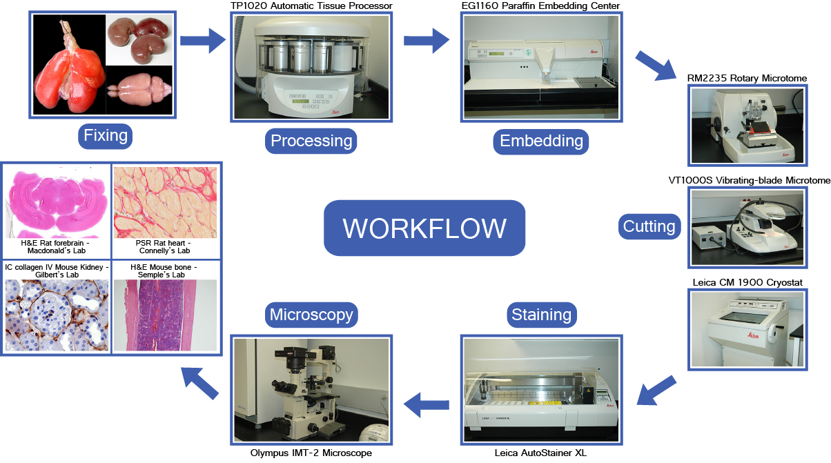
Histology is the study of the microscopic structure of tissues and cellular morphology, utilizing specialized staining protocols in conjunction with light and electron microscopy. Typical histological preparation comprises four main steps: fixation, processing/embedding, sectioning, and staining.
The Histology Core is equipped with a comprehensive range of histology instruments and offers technical expertise for both automated and manual processing of fresh or fixed tissue samples. These services enable detailed investigation of tissue structure, function, and disease progression across various biological systems and animal models.
Equipment
Training
All new users must attend a separate training session for each type of equipment. Please contact the Specialist for more information.
QUICK START GUIDES
HistoCORE Pegasus Tissue Processor – Quick Start Guide
Epredia PrintMate AS Cassette Printer – Quick Start Guide
Epredia Slidemate Slide Printer – Quick Start Guide
Leica HistoCore Arcadia Embedding Cenre – Quick Start Guide
Resources
If you are a new user, require access, or require refresher training, please contact the Specialist. See below for general RCF resources.
Histology Workflow
EXTERNAL INVESTIGATOR RESOURCES
We offer external services at a cost depending on the scope of the work. Please contact us for more information.
Contact Us
For inquiries, contact Xiaofeng Lu at Xiaofeng.Lu@unityhealth.to
Click here to view all RCF specialists.