Immunofluorescence (IF): Detection of a target using an antibody and subsequent visualization using a fluorophore.
Immunofluorescence (IF) includes imunohistofluorescence (IHF) and Immunocytofluorescence (ICF).
Imunohistofluorescence (IHF) detects a target in tissue using an antibody and subsequent visualization using a fluorophore; and Immunocytofluorescence (ICF) detects a target in cells using an antibody and subsequent visualization using a fluorophore.
The basic approach of using antibodies to bind antigens is the same. However, the way to determine the location of the antibodies differs.
Immunofluorescent methods use antibodies labeled with fluorescent fluorophores. The fluorochrome is detected by fluorescence microscopy, always involving excitation of fluorescence with a defined wavelength band of light and detection of the longer-wave emission from the fluorochrome.
Immunohistochemical methods encompass a broad range of chemical reactions used to produce a microscopically visible signal, usually by producing a stain or sometimes emitting photons.
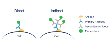
Direct IF uses a single antibody directed against the target of interest. The primary antibody is directly conjugated to a fluorophore.
Indirect IF uses two antibodies. The primary antibody is unconjugated and a fluorophore-conjugated secondary antibody directed against the primary antibody is used for detection.