The aim of Tissue Processing is to remove water from tissues and replace with a medium that solidifies to allow thin sections to be cut. Biological tissue must be supported in a hard matrix to allow sufficiently thin sections to be cut, typically 5 – 6 μm. For light microscopy, paraffin wax is most frequently used. The main steps in this process are dehydration, clearing and filtration.
Wet fixed tissues (in aqueous solutions) cannot be directly infiltrated with paraffin. Water from the tissues must be removed by dehydration. This is usually done with a series of alcohols, 70% to 95% to 100%.
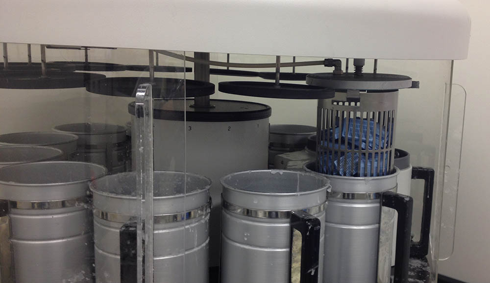
Although the tissue is now essentially water-free, we still cannot infiltrate it with wax because wax and ethanol are largely immiscible. We therefore have to use an intermediate solvent that is fully miscible with both ethanol and paraffin wax. This solvent will displace the ethanol in the tissue, and then this in turn will be displaced by molten paraffin wax. The most common clearing agent is xylene.
Tissue can now be infiltrated with a suitable histological wax. Although many different reagents have been evaluated and used for this purpose over many years, the paraffin wax-based histological waxes are the most popular.
Sectioning is the process of cutting thin slices of a sample from an embedding block using a microtome. The sections are usually on the order of 4-10 µm thick for use with light microscopy.
To cut sections, a metal, glass, or diamond blade is first secured on a microtome. The sample is then placed in a sample holder. Next, the sample is advanced to the cutting surface and drawn across the blade to create a slice of desired thickness.
Following sectioning, samples are placed on slides. For paraffin embedded samples, slices are first placed into a warmed water bath and are then lifted out of the water onto the slide and allowed to dry.
This is the most common microtomes used to cut paraffin blocks in the labs. This device operates with a staged rotary action such that the actual cutting is part of the rotary motion. The typical cut thickness for a rotary microtome is between 1 and 50 µm.
The vibrating microtome operates by cutting using a vibrating blade, allowing the resultant cut to be made with less pressure than would be required for a stationary blade. The cut thickness is usually around 50–500 µm for live tissue and 10–500 µm for fixed tissue.
Cryostat is used to cut frozen sections at very low temperatures (-20 -30 C). The cryostat is just a refrigerated box containing a microtome with an external wheel for rotating the microtome. The section thickness range is 1 – 100 µm.