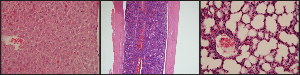
Hematoxylin and eosin stain (H&E) is one of the principal stains in histology. This stain has been unchanged for many years because it works well with a variety of fixatives and displays a broad range of cytoplasmic, nuclear, and extracellular matrix features. So far it is still the most widely used stain in medical diagnosis and is often the gold standard.
Hematoxylin is a compound extracted from the heartwood of the logwood tree. Haematoxylin can be considered as a basic dye. It is used to stain acidic structures a purplish blue. DNA in the nucleus, and RNA in ribosomes and in the rough endoplasmic reticulum are both acidic, and so haemotoxylin binds to them and stains them purple. Haematoxylin alone is not technically a dye, and will not directly stain tissues. It therefore needs to be used in combination with a “mordant” – a compound that helps it link to the tissue to form a tissue-mordant-haematoxylin linkage. The mordant used is typically a metal cation, such as aluminium.
Eosin is anionic and acts as an acidic dye. It is negatively charged and stains basic (or acidophilic) structures red or pink. Most proteins in the cytoplasm are basic, and so eosin binds to these proteins and stains them pink. This includes cytoplasmic filaments in muscle cells, intracellular membranes, and extracellular fibres.
As its name suggests, H&E stain makes use of a combination of two dyes – haematoxylin and eosin. Tissue stained with haematoxylin and eosin shows cytoplasm stained pink-orange and nuclei stained darkly, either blue or purple. Eosin also stains red blood cells intensely red.
Progressive staining – When tissue is left in the stain just long enough to reach the proper end point. The slides have to be examined at different interval to find out when the staining is optimum.
Regressive staining – In this method the tissue is overstained and then destained (differentiate) until the proper endpoint is reached.