Pre-Clinical Imaging

The Research Facilities provides access to in vivo imaging equipment.

Online scheduling is available only to those individuals who have received training on equipment. Review the Imaging guideline on how to receive training. 


Acquisition Systems:

In Vivo Imaging Equipment:

Whole animal photo-acoustic imaging of anesthetized mice. Capable of longitudinal studies done on the same animal.  Allows for the real time measurement of both deoxygenated and oxygenated hemoglobin without the requirement of labels. In addition, dyes (such as ICG) may used and spectrally unmixed from hemoglobin in post processing steps.
""
Heidleberg Spectralis (HRA+OCT)
Simultaneous fundus (retina, optic disc, macula, fovea, posterior pole) imaging and optical coherence tomography. Adapted for use on animals. Imaging Modalities: OCT, infrared, autoflourescence, fluorescein angiography, ICG angiography, red free photography. Contact Dr. Yucel for more information: Yeni.Yucel@unityhealth.to

""

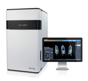
The Newton 7.0 FT500 is capable of both bioluminescence (2D and 3D tomography) as well as fluorescence imaging.

Designed for preclinical in vivo imaging of small animals with a fully automated heated stage and an integrated anesthesia system – allowing for up to 5 mice to be imaged simultaneously. (Also capable of ex vivo and in vitro applications.)

Camera – wide aperture (f/0.7) 16-bit cooled CCD camera

Fluorescence – 8 LED excitation channels, and 8 narrow bandpass emission filters for a wide array of fluorophores (420nm – 780nm).

Image of NEWTON 7.0
See Bioimaging Equipment for more information.

MILabs’ U-CT is available for use as a specialty service. Click here for more information.